Advanced Imaging Techniques are crucial in orthopaedics for diagnosing and treating musculoskeletal conditions. These imaging tools provide detailed views of bones, joints, and soft tissues, enabling accurate diagnosis and effective treatment planning. This article explores various advanced imaging techniques, focusing on the use of X-rays, MRI advancements, and comparisons between these techniques.
Which of the Following Advanced Imaging Techniques Uses X-rays?
Among advanced imaging techniques, several utilize X-rays. These include:
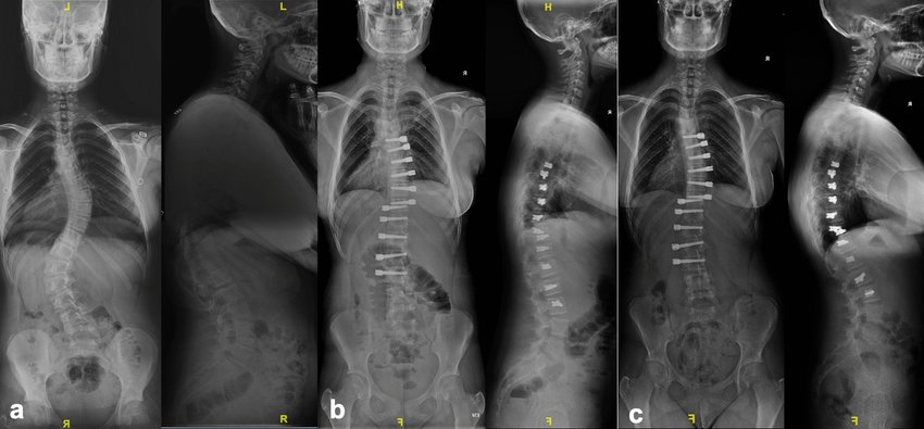
- Computed Tomography (CT) Scans: CT scans use X-rays to create detailed cross-sectional images of the body. They provide more detailed views than standard X-rays, making them useful for diagnosing complex bone fractures and other musculoskeletal issues.
- Fluoroscopy: This technique involves continuous X-ray imaging to visualize the movement of bones and joints in real-time. It is often used during orthopedic procedures to guide surgical instruments.
- Digital X-rays: Digital X-rays are a modern form of traditional X-rays. They use digital sensors instead of film to capture images, offering improved clarity and faster processing.
What Are the Advanced Imaging Techniques in Radiology?
Radiology encompasses various advanced imaging techniques used to diagnose and evaluate bone and joint conditions. Key techniques include:
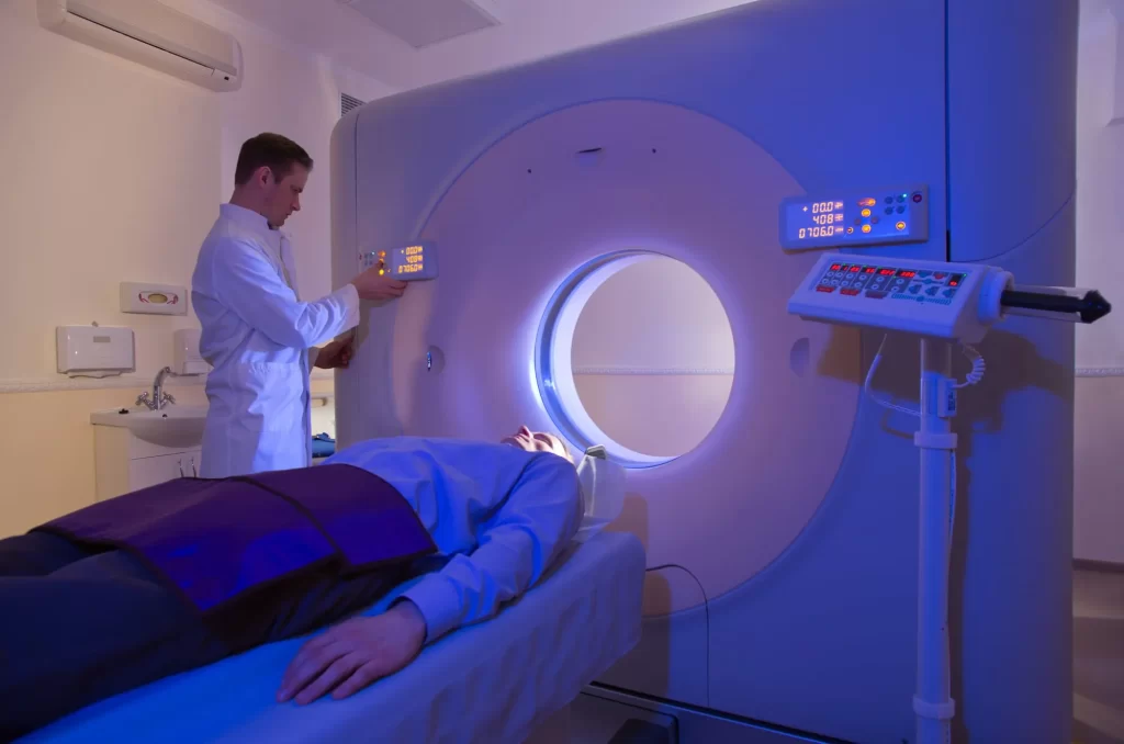
- Magnetic Resonance Imaging (MRI): MRI uses strong magnetic fields and radio waves to create detailed images of soft tissues, including muscles, tendons, and cartilage. It is invaluable for diagnosing injuries and conditions affecting these tissues.
- Computed Tomography (CT) Scans: As mentioned, CT scans provide detailed cross-sectional images using X-rays. They are particularly useful for visualizing complex fractures and assessing bone alignment.
- Ultrasound: This imaging technique uses high-frequency sound waves to create images of soft tissues and fluid-filled areas. It is commonly used for assessing muscle injuries, tendon conditions, and joint effusions.
- Positron Emission Tomography (PET) Scans: PET scans use radioactive tracers to detect metabolic activity in tissues. While less common in orthopaedics, PET scans can help identify bone infections or tumors.
- Bone Scintigraphy (Bone Scan): This technique involves injecting a small amount of radioactive material to highlight areas of abnormal bone activity. It is useful for detecting bone infections, tumors, and stress fractures.
What Is Common Between X-ray and MRI Techniques?
Despite their different technologies, X-ray and MRI techniques share some common aspects:
- Diagnostic Purpose: Both X-rays and MRIs aim to diagnose musculoskeletal conditions and injuries. They provide detailed images that help physicians make informed decisions about treatment.
- Non-invasive: Both imaging techniques are non-invasive, meaning they do not require surgery or insertion of instruments into the body. They offer a way to visualize internal structures without physical intervention.
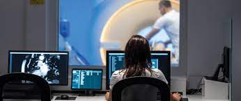
- Image Interpretation: Radiologists interpret the images produced by X-rays and MRIs. Their expertise is crucial for accurately diagnosing conditions based on the imaging results.
- Patient Preparation: Both techniques may require some level of patient preparation. For X-rays, patients might need to remove metal objects, while MRI patients must stay still and may need to remove certain items.
What Is the Advanced Technique of MRI?
Recent advancements in MRI technology include:
- Functional MRI (fMRI): fMRI measures brain activity by detecting changes in blood flow. Although primarily used in neurology, it has applications in orthopaedics for assessing brain responses related to pain or injury.
- High-Resolution MRI: This technique provides more detailed images by using higher magnetic field strengths and advanced imaging sequences. It is particularly useful for visualizing small structures and subtle injuries.
- 3D MRI Imaging: 3D MRI creates three-dimensional images from multiple two-dimensional slices. This advanced technique offers a comprehensive view of anatomical structures, aiding in precise diagnosis and surgical planning.
- Magnetic Resonance Arthrography (MRA): MRA involves injecting a contrast agent into a joint before performing an MRI. This technique enhances the visualization of joint structures, such as cartilage and ligaments, making it useful for diagnosing complex joint issues.
Conclusion
Advanced imaging techniques, including X-rays and MRIs, play a pivotal role in orthopaedics by providing detailed insights into musculoskeletal conditions. X-rays, through techniques like CT scans and fluoroscopy, offer valuable diagnostic information. MRI advancements, such as functional MRI and high-resolution imaging, provide enhanced views of soft tissues and complex injuries.
Understanding these imaging techniques helps healthcare professionals make informed decisions about diagnosis and treatment. By leveraging the strengths of each technique, orthopaedic specialists can deliver precise and effective care, improving patient outcomes and facilitating optimal recovery.